 Deutsch
 English
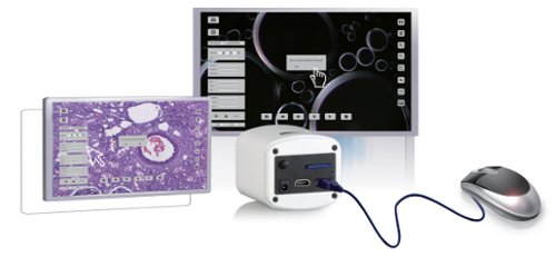
HDMI/USB/Network Microscope Camera DF5
The HDC DF5 microscope camera is the right choice for users who have very high quality demands for image quality on one hand and want to remain flexible regarding system compatibility on the other. The excellent image quality is guaranteed by the built-in SONY IMX585(C) CMOS color image sensor with a size of 1/1.2" (11.14x6.26 mm). This allows for recording in a resolution of 3840 x 2160. In terms of compatibility, this microscope camera can be operated with or without a PC and used in various modes: HDMI (without PC), USB (with Windows PC), or Network mode (with Windows PC).
The most important functions of the camera are listed below - functions marked with (*) are only available when using the included Windows software - and not in pure HDMI mode.
Multi-Signal Output: The camera can be connected either directly to an HDMI monitor (via the included HDMI cable) or alternatively to a PC (via the included USB cable). In both modes, the camera provides sufficiently high frame rates to track and record microscopic movements. The camera can also be used in mixed operation, where you work on the PC and simultaneously transmit the image via the HDMI output to a second HDMI monitor.
Video Stream over Network: The HDC DF5 microscope camera can be connected to the local network via the included network cable and stream the live image over the network. PCs on the same network (assuming the same network segment and the installed Windows software application for the camera) will detect the camera, and the user can receive the video stream over the network instead of via USB. This allows you to provide the live image to colleagues who are not present in the same room.
Flexible Exposure Times: In addition to auto-exposure, you can also manually set the exposure time and gain depending on the application. The exposure time can be set between 0.048~1000 msec.
ROI - Flexible White Balance: You can choose between an automatic and a manual white balance. Additionally, you can activate an automatic white balance with reference to a selectable image region (ROI - region of interest). In the ROI white balance mode, you frame a rectangular area on the image (the rectangle can be moved and its size adjusted with the mouse). The software then uses only this area as a reference for white.
Auto-Stitch*: Multiple partially overlapping images can be combined by the software at the touch of a button into a single overall image. The sample image below was generated automatically from five partial shots; the individual images had an overlap of approximately 10-20% with each other during capture.
Extended Depth of Field* (EDF): Sometimes it is not possible to capture the entire area in focus because the depth of field is limited by the optics. After capturing multiple images with different focus settings, the software generates a composite image using stacking, taking only the sharp areas from each individual image. The sample image below/right was generated automatically from four partial shots. The partial shots contained only partially sharp areas, as can be seen, for example, in the left partial image.
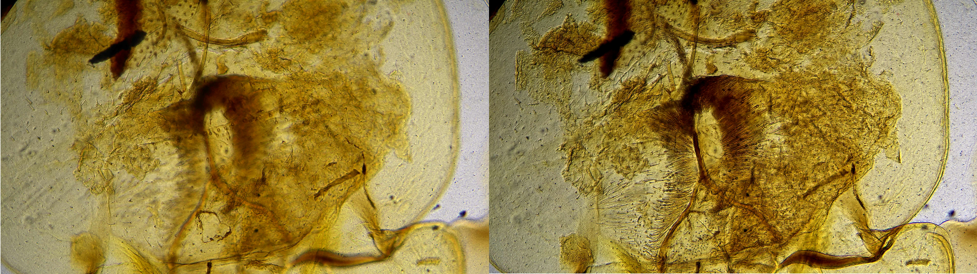
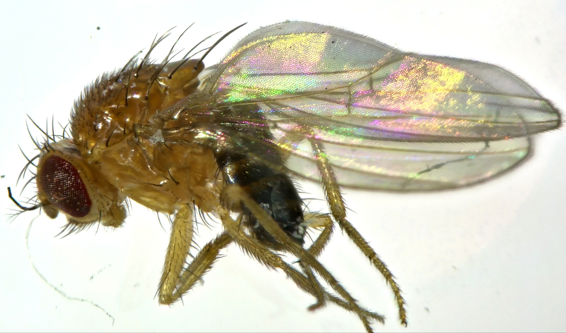
In the example below, the EDOF function was used to render most of the insect in sharp focus.
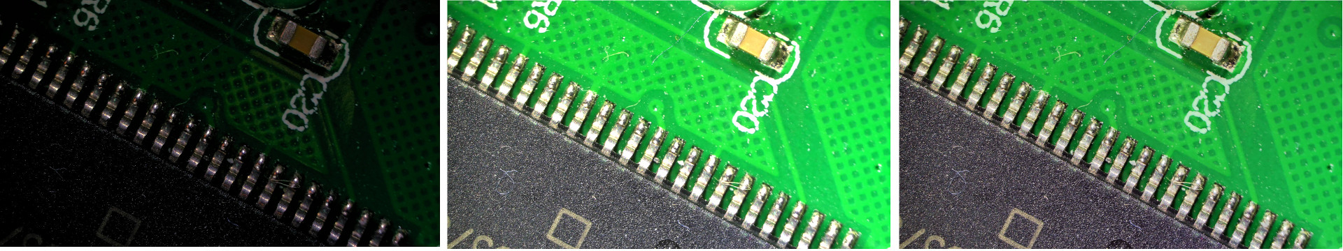
HDR - High Dynamic Range*: This function merges a sequence of differently exposed images into one image with a higher dynamic range. In the example below, we created a new image from two shots, one underexposed and one overexposed, resulting in ideal exposure for each image area.
Zoom – Mirror - Compare - Freeze: Using the software, you can zoom in and out of the image and mirror the image horizontally/vertically. Furthermore, you can split the screen vertically into two halves: the left half shows the live image and the right half shows an existing image from your gallery to demonstrate specimen comparisons. Additionally, you can freeze the live image and then release it for viewing again.
Measurements: The camera software can perform measurements after user calibration has been performed.
Time-Lapse*: Interval recordings can be programmed, where the time interval between shots and the maximum number are configurable. This allows, for example, biological decay processes to be documented over time.
Built-in Mouse Control (HDMI mode): The included mouse is connected to the USB port of the camera. With the mouse, you can then operate the On-Screen Display menu and use it to capture images and videos and adjust all camera settings - completely without a PC and software installations!
Video Recording on Removable Storage Media (HDMI mode): Videos/Images can be recorded directly in 4K quality onto a removable SD card or USB stick.
Built-In Player: (HDMI mode): Already captured images and videos are stored on the camera's removable storage media. You can browse through them and have the camera play them back on the monitor – completely without a PC!
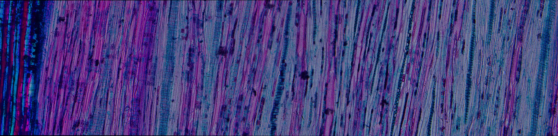
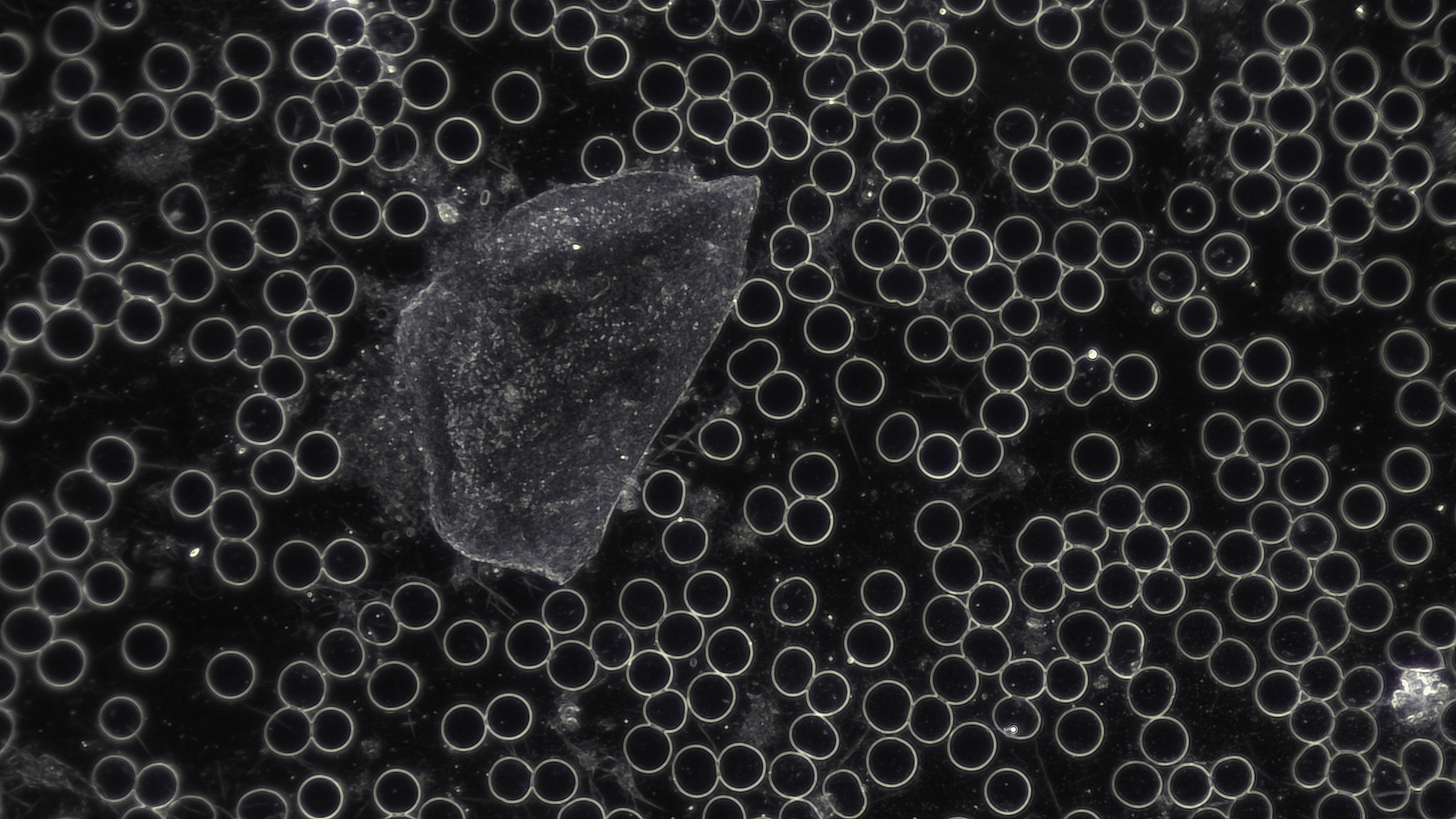
Sample Recordings
In the following images, you can see sample recordings from the HDC DF5 camera in dark field mode.

In the following video, you can see a video recording from the HDC DF5 (Sample: Blood drop, Mode: Dark field, Magnification: 400X, Microscope: OPTIKA B-510DK). The recording was made in HDMI mode (recorded directly to the removable storage medium of the microscope camera).
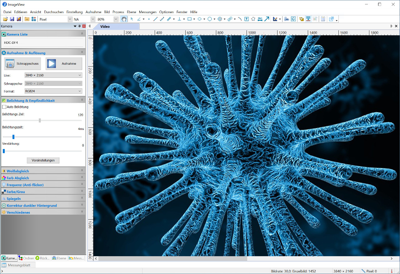
Windows User Interface
The multilingual interface of the included Windows software provides, on one hand, control options for the camera parameters (exposure, gain, capture) and, on the other hand, options for image correction (white balance, color tones, etc.) as well as access to the advanced functions (measurement, extended depth of field, auto-stitch, etc.)
Technical Specifications
- Sensor: Sony IMX585(C), Size: 1/1.2"
- Pixel Size: 2.9 x 2.9 micrometers
- Sensor Output: 60fps at 3840×2160
- Frame Rate (HDMI, USB 3.0, Ethernet): 60, 30, 30 fps
- Storage Formats: Video 8MP (3840 x 2160) H264/H265 MP4, Single Image: 8MP (3840 x 2160) JPEG/TIFF/RAW
- Exposure Time (msec): 0.048 - 1000
- White Balance: manual, automatic, region of interest
- Language Support ImageView Windows Software: German, English, French, Italian, Spanish, etc.
- Special Functions of the ImageView Software: Merging of images (auto-stitch), Extended Depth of Field, HDR, Segmentation, Time-Lapse, Measurements
- Outputs: HDMI, USB, Ethernet, removable storage media
- Compatibility: Windows 10/11 (32/64 Bit)
- System Requirements: CPU Intel Core2 2.8 GHz or better, min. 4GB RAM
- Mount: C-Mount
- Weight (g): 550 without accessories
- Dimensions (mm): 98.3 x 78 x 65
- Technical Datasheet (PDF): Download under the "Media" tab
Scope of Delivery
- Microscope Camera
- Power Supply AC 100-240V 50/60 Hz, DC 12V 1A, EU Standard
- ImageView Software for Windows
- HDMI Cable
- USB 3.0 Cable (A-male to A-male), 2.0 m
- USB Mouse
- Camera Manual (German): Download under the "Media" tab
- Software Manual (English)
- Software (on USB Memory Stick)
Manufacturer's Warranty
- 18 months from date of purchase (EU)